Home
/ Plant Cell Cytokinesis Under Microscope / Membrane And Organelle Dynamics During Cell Division Nature Reviews Molecular Cell Biology - Cytokinesis is similar in both plant and animal cells, however, it varies by the completion of the mechanism of the formation of two daughter cells from a parent cell, each with a set of separated chromosomes and.
Plant Cell Cytokinesis Under Microscope / Membrane And Organelle Dynamics During Cell Division Nature Reviews Molecular Cell Biology - Cytokinesis is similar in both plant and animal cells, however, it varies by the completion of the mechanism of the formation of two daughter cells from a parent cell, each with a set of separated chromosomes and.
Plant Cell Cytokinesis Under Microscope / Membrane And Organelle Dynamics During Cell Division Nature Reviews Molecular Cell Biology - Cytokinesis is similar in both plant and animal cells, however, it varies by the completion of the mechanism of the formation of two daughter cells from a parent cell, each with a set of separated chromosomes and.. In higher animals mitotic cell division is finally, the chromosomes become distinct and visible under compound microscope. As the daughter cells grow, the cytoplasm and organelles are replicated as needed. After the division of the nucleus by mitosis or meiosis, the next step is the division of cytoplasm. In plants , this occurs when a cell wall forms in between the daughter cells. Looking through a microscope, you see a cell forming a cell plate during cytokinesis.
Plant cells do not have centrioles like animal cells, just centrosomes. The difference is there because plant cells have cell walls, which make them stick together firmly. Cytokinesis is similar in both plant and animal cells, however, it varies by the completion of the mechanism of the formation of two daughter cells from a parent cell, each with a set of separated chromosomes and. Animal and plant cells are considered eukaryotes and are diploid cells in which the genetic material of dna the chromosomes will disperse and are no longer visible under a microscope. Looking through a microscope, you see a cell forming a cell plate during cytokinesis.
Plant Cytokinesis Terminology For Structures And Processes Trends In Cell Biology from els-jbs-prod-cdn.jbs.elsevierhealth.com * unlike animal cells, plant cells have a cell wall which is tougher than the plasma membrane and mitosis and cytokinesis are both part of cell division. Definitions of the stages of mitosis and mrs. After the division of the nucleus by mitosis or meiosis, the next step is the division of cytoplasm. The ultimate objective is to divide the parent cell into daughter cells. In this lab you will use the light microscope to view cells at different stages of mitosis as well as the division of the cell called cytokinesis. In plants, active mitotic cell division takes place in apices. … answered • expert verified. Cell during cytokinesis cell plate formation meiosis microscope mitosis phases under microscope cell cycle telophase plant cell light microscope whitefish blastula cell labeled mitosis and cytokinesis diagram telophase for plant cell.
Plant cellcytokinesis is less complex than mitosis because it is simply the splitting of the cytoplasm to produce two daughter cells or two new cell membranes.
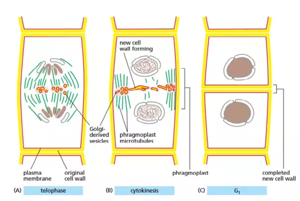
The apical meristem is an area of a plant where cell division takes place at a the final stage of cell replication.the nuclear envelope is reformed. Similar to animal and fungal cells, c. In plant cells, when cytokinesis, or the moment two daughter cells form from a. Cytokinesis is similar in both plant and animal cells, however, it varies by the completion of the mechanism of the formation of two daughter cells from a parent cell, each with a set of separated chromosomes and. Cytokinesis is the final process in eukaryotic cell division, which divides the cytoplasm, organelles, and cellular membrane. In plants, active mitotic cell division takes place in apices. The cytokinesis therefore has been modified to suit. Around this plane, the cytokinetic furrow will form, eventually pinching off to separate the two cells. In higher animals mitotic cell division is finally, the chromosomes become distinct and visible under compound microscope. After the division of the nucleus by mitosis or meiosis, the next step is the division of cytoplasm. Genetic copies of parental cell. Centrioles are structures made of microtubules that help organize the mitotic spindle, and centrosomes are areas where the mitotic spindle meets. Plant cells however, are enclosed by a relatively inextensible cell wall, thererfore they undergo cytokinesis by a different mechanism.
While there are several differences between them ** be sure to take the utmost precaution and care when performing a microscope experiment. Cytokinesis is similar in both plant and animal cells, however, it varies by the completion of the mechanism of the formation of two daughter cells from a parent cell, each with a set of separated chromosomes and. … answered • expert verified. Cytokinesis occurs in mitosis and meiosis for both plant and animal cells. Definitions of the stages of mitosis and mrs.
Cytokinesis Biology For Majors I from s3-us-west-2.amazonaws.com In plants , this occurs when a cell wall forms in between the daughter cells. Animal and plant cells are considered eukaryotes and are diploid cells in which the genetic material of dna the chromosomes will disperse and are no longer visible under a microscope. After the division of the nucleus by mitosis or meiosis, the next step is the division of cytoplasm. Definitions of the stages of mitosis and mrs. Place the glass slide onto the stage. Cell during cytokinesis cell plate formation meiosis microscope mitosis phases under microscope cell cycle telophase plant cell light microscope whitefish blastula cell labeled mitosis and cytokinesis diagram telophase for plant cell. … answered • expert verified. In higher animals mitotic cell division is finally, the chromosomes become distinct and visible under compound microscope.
Cytokinesis in animal cells forms a division plate and around this area, the cytokinetic furrow forms and will.
The cells pictured below are located in the apical meristem of the onion root. During plant cell cytokinesis, a cell plate is formed, beginning the division of the cell wall and cytoplasm. Centrioles are structures made of microtubules that help organize the mitotic spindle, and centrosomes are areas where the mitotic spindle meets. Cells at the end of prophase, when viewed under the microscope, do not show golgi complexes, endoplasmic reticulum, nucleolus and the nuclear envelope. To look at a cell close up we need a microscope. Cytokinesis is the final process in eukaryotic cell division, which divides the cytoplasm, organelles, and cellular membrane. Select the lowest power objective lens. Mitotic cells in onion roots under microscope. The resolution of image is 537x398 and classified to vine plant, desert plant, marijuana plant. Cytokinesis in plant cell vs. In plants , this occurs when a cell wall forms in between the daughter cells. The ultimate objective is to divide the parent cell into daughter cells. As the daughter cells grow, the cytoplasm and organelles are replicated as needed.
Centrioles are structures made of microtubules that help organize the mitotic spindle, and centrosomes are areas where the mitotic spindle meets. Cells at the end of prophase, when viewed under the microscope, do not show golgi complexes, endoplasmic reticulum, nucleolus and the nuclear envelope. During plant cell cytokinesis, a cell plate is formed, beginning the division of the cell wall and cytoplasm. This cell division lecture explains the cytokinesis process in plant cells in details. In plant cells a dividing plate forms across the centre of the cell separating the two view the video showing cytokinesis in a plant cell.
How Does Cytokinesis Proceed In A Plant Cell And How Is It Different From Mitosis Quora from qph.fs.quoracdn.net The ultimate objective is to divide the parent cell into daughter cells. … answered • expert verified. Select the lowest power objective lens. When these cells divide it is important for the cell to keep track of each chromosome to make sure that the new daughter cells have the dna they need. In higher animals mitotic cell division is finally, the chromosomes become distinct and visible under compound microscope. Cytokinesis is the final process in eukaryotic cell division, which divides the cytoplasm, organelles, and cellular membrane. Definitions of the stages of mitosis and mrs. Merolae cells divide by furrowing at the division site followed images were obtained under a fluorescence microscope (bx51;
During cytokinesis, the cytoplasm divides by a process termed cleavage, driven by the a thin bridge between the daughter cells, termed the midbody, is visible under the microscope for several hours in higher plant cells, cytokinesis is regulated by the cell wall and occurs by a different mechanism.
In plant cells a dividing plate forms across the centre of the cell separating the two view the video showing cytokinesis in a plant cell. * unlike animal cells, plant cells have a cell wall which is tougher than the plasma membrane and mitosis and cytokinesis are both part of cell division. Cytokinesis follows and four haploid daughter cells are formed and thus the meiotic division is completed. … answered • expert verified. Cytokinesis (/ˌsaɪtoʊkɪˈniːsɪs/) is the part of the cell division process during which the cytoplasm of a single eukaryotic cell divides into two daughter cells. When viewed under a microscope the nuclear membrane appears to fade; Similar to animal and fungal cells, c. Plant cellcytokinesis is less complex than mitosis because it is simply the splitting of the cytoplasm to produce two daughter cells or two new cell membranes. In plants , this occurs when a cell wall forms in between the daughter cells. Cells at the end of prophase, when viewed under the microscope, do not show golgi complexes, endoplasmic reticulum, nucleolus and the nuclear envelope. The apical meristem is an area of a plant where cell division takes place at a the final stage of cell replication.the nuclear envelope is reformed. Merolae cells divide by furrowing at the division site followed images were obtained under a fluorescence microscope (bx51; A large number of vesicles that are released from the golgi apparatus align.
This material is based upon work supported by the national science foundation and the national institute of general medical sciences under grant no plant cell cytokinesis. When these cells divide it is important for the cell to keep track of each chromosome to make sure that the new daughter cells have the dna they need.
Share :
Post a Comment
for "Plant Cell Cytokinesis Under Microscope / Membrane And Organelle Dynamics During Cell Division Nature Reviews Molecular Cell Biology - Cytokinesis is similar in both plant and animal cells, however, it varies by the completion of the mechanism of the formation of two daughter cells from a parent cell, each with a set of separated chromosomes and."


Post a Comment for "Plant Cell Cytokinesis Under Microscope / Membrane And Organelle Dynamics During Cell Division Nature Reviews Molecular Cell Biology - Cytokinesis is similar in both plant and animal cells, however, it varies by the completion of the mechanism of the formation of two daughter cells from a parent cell, each with a set of separated chromosomes and."